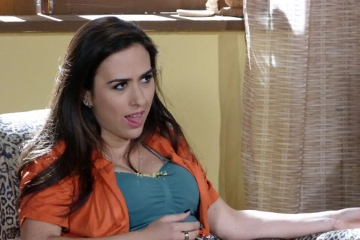
The subcellular localisation of trypanosome RRP6 and its association with the exosome (2023)

Molecular and Biochemical Parasitology

Volume 151, Issue 1,

January 2007

, Pages 52-58

Author links open overlay panel, , ,

Abstract

The exosome, a complex of 3′-exoribonucleases and associated proteins, is involved in the degradation of eukaryotic mRNAs in the cytoplasm, and has RNA processing and quality control functions in the nucleus. In yeast, the nuclear exosome differs from the cytoplasmic one in that it contains an additional non-essential component, Rrp6p. In contrast, a small proportion of human RRP6 has been shown to localise to the cytoplasm as well. When we purified the Trypanosoma brucei exosome from cytosolic extracts we found RRP6, apparently in stoichiometric amounts. We here confirm that RRP6 is in the trypanosome cytoplasm and nucleus. The level of RRP6 was unaffected by depletion of core exosome subunits by RNA interference and over-expression of tagged RRP6 was possible, indicating that RRP6 can be present independent of exosome association.

Introduction

The exosome is a protein complex which is involved in 3′→5′ degradation of diverse RNAs. It is composed of a “core” of six subunits related to Escherichia coli RNase PH, several of which have been shown to have 3′→5′ exonuclease activity, and three subunits with S1 domains, and is present in yeast, trypanosomes, animals and plants [1], [2], [3], [4], [5].

The exosome has multiple functions in the nucleus, including processing of rRNAs and small nucleolar (sno) RNAs, and quality control of mRNAs (reviewed in [6] and see [7], [8], [9], [10], [11]). In the cytoplasm, it is involved in the degradation of both normal and defective mRNAs [12], [13], [14], [15], [16], [17], [18], [19], [20], [21]. By analogy with the structure of prokaryotic polynucleotide phosphorylase, Aloy et al. [22] suggested that the six RNase PH subunits of the exosome form a ring, with the three S1 domain subunits balanced on top. This concept has been supported by analyses using yeast and mammalian two-hybrid systems [23], [24] and using mass spectrometry [25]. The archaeon Sulfolobus solfataricus has an exosome-like complex [26] in which three copies each of two RNase PH subunits form a hexameric core structure [27]. In yeast, all of the RNase PH and S1 domain subunits are essential for viability [2].

Various other proteins are associated with the yeast and human exosomes, often in sub-stoichiometric amounts. One of these sub-stoichiometric components is RRP6, an RNase D enzyme. S. cerevisiae Rrp6p is found only in the nucleus [2], where it is involved in multiple processes, including linkage of transcription with RNA processing [28]. Deletion of the gene encoding Rrp6p causes lethality at 37°C, but not at 30°C [2]. In Drosophila tissue culture cells, the Rrp6p homologue is predominantly, but not exclusively, nuclear [29]. The human Rrp6p is called PM/Scl-100, a target of auto-antibodies in patients with polymyositis-scleroderma overlap syndrome. Pm/Scl-100 was initially found to be undetectable in a cytoplasmic extract using the human autoimmune sera [2], but a later report showed that a small quantity was cytoplasmic [3]. In support of a partially cytoplasmic location for PM/Scl-100, depletion of the subunit using siRNA inhibited decay of an unstable mRNA containing a nonsense codon [14].

We have previously shown that the Trypanosoma brucei exosome is similar to those of other eukaryotes [30]. There are six RNase PH subunits (EAP1, EAP2, EAP4, RRP41A, RRP41B, RRP45), and three S1 domain subunits (RRP40, CSL4, RRP4). In addition, however, the RRP6 RNase D, and an additional small subunit called EAP3 were present in apparently stoichiometric amounts. RNA-interference (RNAi)-mediated depletion of each of the individual exosome components, including RRP6, in procyclic trypanosomes (the life-cycle stage which multiplies in the Tsetse fly) inhibited growth and 5.8 S rRNA processing [30], [31]. Depletion of the core subunit RRP45 also inhibited growth, and degradation of unstable mRNAs, in bloodstream forms [32]. Analyses using the yeast two-hybrid system gave structural predictions consistent with those from yeast and mammals; in addition, an interaction was found between trypanosome RRP6 and EAP3. The effects of genetic manipulation yielded more insights into exosome structure. First, over-expression of tagged version of either RRP4 (and S1 domain subunit) or RRP45 (a core subunit) resulted in a decrease in abundance of the endogenous, untagged protein, suggesting that both RRP4 and RRP45 are unstable when not associated with the exosome complex. Second, depletion of the core subunits RRP45, RRP41B, EAP2 and EAP4 caused co-depletion of RRP4 and RRP45, suggesting that the absence of any of these subunits destabilises the entire complex. More surprisingly, depletion of RRP6 and EAP3 also caused decreases in RRP4 and RRP45 [31]. This result suggested that – in dramatic contrast with the situation in yeast – RRP6 might either be a central, part of the exosome structure, or be involved in exosome assembly. This paper describes the results of further experiments to define the location and functional role of RRP6.

Section snippets

Trypanosomes

Procyclic forms of T. brucei expressing the tet repressor were cultured as described [33]. Cell lines harboring RNAi plasmids targeting RRP44 or each exosome components, or expressing TAP-tagged RRP4 or RRP45, were grown and induced as described [30], [31].

Expression of TbRRP6 in E. coli and generation of antiserum

Several attempts to produce full-length RRP6 in E. coli failed, so instead, a fragment of the TbRRP6 open reading frame corresponding to amino acids 468–588 was amplified using primers CZ1667 (CTC GGA TCC ATG TCT GCG GTT AAG) and CZ1668 (CAC

TbRRP6 is stable in the absence of other exosome subunits

We had previously shown that depletion of RRP6 results in a decrease in the amounts of both RRP45 and RRP4. We had, not, however, been able to determine the fate of RRP6 after depletion of other exosome subunits. To find out of exosome depletion affected RRP6, we made lysates of procyclic trypanosomes in which expression of individual exosome subunits had been reduced by RNAi, and measured RRP6 levels using a new specific antiserum (see Section 2 and Fig. 3A for details). The results from this

Discussion

We have previously shown that in trypanosomes, RRP6 is essential for maintaining the structural integrity of the exosome. Results presented here show that in contrast, a reduction in the amount of exosome does not affect the abundance of RRP6. The core exosome components RRP45, RRP41B, EAP2 and EAP4 are essential for exosome integrity [31] but RRP6 levels were not reduced by depletion of any of these subunits (Fig. 1) even though the migration of the protein in glycerol gradients was clearly

Acknowledgements

This work was supported by the Deutsche Forschungsgemeinshaft, CL112/7 and Cl112/9, and Graduiertenkolleg 300. We thank Noreen Williams and Marilyn Parsons for antibodies. The experiments shown in Fig. 1, Fig. 2 were done by SH under the supervision of AE, and those in Fig. 3 by SH, AE, and MC. MC also did the TAP-tagging and mutagenesis supervised by CC. CC wrote the grant applications and all authors contributed to writing the manuscript.

References (39)

  • P. Mitchell et al.

    The exosome: a conserved eukaryotic RNA processing complex containing multiple 3′-5′ exoribonucleases

    Cell

    (1997)

  • R. Brouwer et al.

    Three novel components of the human exosome

    J Biol Chem

    (2001)

  • R. Raijmakers et al.

    The exosome, a molecular machine for controlled RNA degradation in both nucleus and cytoplasm

    Eur J Cell Biol

    (2004)

  • S. West et al.

    Adenylation and exosome-mediated degradation of cotranscriptionally cleaved pre-messenger RNA in human cells

    Mol Cell

    (2006)

  • L. Vasiljeva et al.

    Nrd1 Interacts with the nuclear exosome for 3′ processing of RNA polymerase II transcripts

    Mol Cell

    (2006)

  • J. LaCava et al.

    RNA degradation by the exosome is promoted by a nuclear polyadenylation complex

    Cell

    (2005)

  • A.J. Carpousis et al.

    mRNA degradation. A tale of poly(A) and multiprotein machines

    Trends Genet

    (1999)

  • C.Y. Chen et al.

    AU binding proteins recruit the exosome to degrade ARE-containing mRNAs

    Cell

    (2001)

  • F. Lejeune et al.

    Nonsense-mediated mRNA decay in mammalian cells involves decapping, deadenylating, and exonucleolytic activities

    Mol Cell

    (2003)

  • P. Mitchell et al.

    An NMD pathway in yeast involving accelerated deadenylation and exosome-mediated 3′→5′ degradation

    Mol Cell

    (2003)

  • R. Raijmakers et al.

    Protein–protein interactions of hCsl4p with other human exosome subunits

    J Mol Biol

    (2002)

  • E. Lorentzen et al.

    Structural basis of 3′ end RNA recognition and exoribonucleolytic cleavage by an exosome RNase PH core

    Mol Cell

    (2005)

  • R. Luna et al.

    Interdependence between transcription and mRNP processing and export, and its impact on genetic stability

    Mol Cell

    (2005)

  • A.M. Estévez et al.

    The roles of inter-subunit interactions in exosome stability

    J Biol Chem

    (2003)

  • S. Biebinger et al.

    Vectors for inducible over-expression of potentially toxic gene products in bloodstream and procyclic Trypanosoma brucei

    Mol Biochem Parasitol

    (1997)

  • J. Zhang et al.

    Developmental regulation of two nuclear RNA binding proteins, p34 and p37, from Trypanosoma brucei

    Mol Biochem Parasitol

    (1998)

  • C. Allmang et al.

    The yeast exosome and human PM-Scl are related complexes of 3′→5′ exonucleases

    Genes Dev

    (1999)

  • E.D. Andrulis et al.

    The RNA processing exosome is linked to elongating RNA polymerase II in Drosophila

    Nature

    (2002)

  • J.A. Chekanova et al.

    Poly(A) tail-dependent exonuclease AtRrp41p from Arabidopsis thaliana rescues 5.8 S rRNA processing and mRNA decay defects of the yeast ski6 mutant and is found in an exosome-sized complex in plant and yeast cells

    J Biol Chem

    (2000)

  • Cited by (37)

    • Stress-induced nuclear depletion of Entamoeba histolytica 3'-5' exoribonuclease EhRrp6 and its role in growth and erythrophagocytosis

      2018, Journal of Biological Chemistry

      The 3′-5′ exoribonuclease Rrp6 is a key enzyme in RNA homeostasis involved in processing and degradation of many stable RNA precursors, aberrant transcripts, and noncoding RNAs. We previously have shown that in the protozoan parasite Entamoeba histolytica, the 5′-external transcribed spacer fragment of pre-rRNA accumulates under serum starvation–induced growth stress. This fragment is a known target of degradation by Rrp6. Here, we computationally and biochemically characterized EhRrp6 and found that it contains the catalytically important EXO and HRDC domains and exhibits exoribonuclease activity with both unstructured and structured RNA substrates, which required the conserved DEDD-Y catalytic-site residues. It lacked the N-terminal PMC2NT domain for binding of the cofactor Rrp47, but could functionally complement the growth defect of a yeast rrp6 mutant. Of note, no Rrp47 homologue was detected in E. histolytica. Immunolocalization studies revealed that EhRrp6 is present both in the nucleus and cytosol of normal E. histolytica cells. However, growth stress induced its complete loss from the nuclei, reversed by proteasome inhibitors. EhRrp6-depleted E. histolytica cells were severely growth restricted, and EhRrp6 overexpression protected the cells against stress, suggesting that EhRrp6 functions as a stress sensor. Importantly EhRrp6 depletion reduced erythrophagocytosis, an important virulence determinant of E. histolytica. This reduction was due to a specific decrease in transcript levels of some phagocytosis-related genes (Ehcabp3 and Ehrho1), whereas expression of other genes (Ehcabp1, Ehcabp6, Ehc2pk, and Eharp2/3) was unaffected. This is the first report of the role of Rrp6 in cell growth and stress responses in a protozoan parasite.

    • Polycistronic trypanosome mRNAs are a target for the exosome

      2016, Molecular and Biochemical Parasitology

      Citation Excerpt :

      In all cells, both N- and C-terminal eYFP fusions of RRP44 and RRP6 were highly concentrated in the nucleus, with a slight enrichment at the edge of the nucleolus, which is here detected by the absence of DAPI staining (Fig. 2A). This localization was similar to the previously published localization of RRP4 [24]. As expected, CBP20 and SmE also localized to the nucleus (Fig. 1B).

      Eukaryotic cells have several mRNA quality control checkpoints to avoid the production of aberrant proteins. Intron-containing mRNAs are actively degraded by the nuclear exosome, prevented from nuclear exit and, if these systems fail, degraded by the cytoplasmic NMD machinery. Trypanosomes have only two introns. However, they process mRNAs from long polycistronic precursors by trans-splicing and polycistronic mRNA molecules frequently arise from any missed splice site. Here, we show that RNAi depletion of the trypanosome exosome, but not of the cytoplasmic 5′-3′ exoribonuclease XRNA or the NMD helicase UPF1, causes accumulation of oligocistronic mRNAs. We have also revisited the localization of the trypanosome exosome by expressing eYFP-fusion proteins of the exosome subunits RRP44 and RRP6. Both proteins are significantly enriched in the nucleus. Together with published data, our data suggest a major nuclear function of the trypanosome exosome in rRNA, snoRNA and mRNA quality control.

    • The DRBD13 RNA binding protein is involved in the insect-stage differentiation process of Trypanosoma brucei

      2015, FEBS Letters

      Citation Excerpt :

      Tetracycline was added to approximately 100 mL of 1313-DRBD13-myc PC trypanosomes growing at a density of 5 × 10⁶ cells/mL. After 3 h of DRBD13-myc induction, the cells were fractionated as described in [26] followed by polysome preparation as in [27]. Western blots were incubated in antibodies against myc tag and small ribosomal subunit protein S9.

      DRBD13 RNA-binding protein (RBP) regulates the abundance of AU-rich element (ARE)-containing transcripts in trypanosomes. Here we show that DRBD13 regulates RBP6, the developmentally critical protein in trypanosomatids. We also show DRBD13-specific regulation of transcripts encoding cell surface coat proteins including GPEET2, variable surface glycoprotein (VSG) and invariant surface glycoprotein (ISG). Accordingly, alteration in DRBD13 levels leads to changes in the target mRNA abundance and parasite morphology. The high consistency of the observed phenotype with known cell membrane exchanges that occur during progression of T. brucei through the insect stage of its life cycle suggests that DRBD13 is an important regulator in this largely unknown developmental process.

    • RNA decay machines: The exosome

      2013, Biochimica et Biophysica Acta - Gene Regulatory Mechanisms

      Citation Excerpt :

      In addition, small quantities are also present in the cytoplasm [34]. A similar localization pattern is observed in Drosophila melanogaster [39] and Trypanosoma brucei [40], suggesting minor cytoplasmic functions of Rrp6 proteins in the corresponding taxa. However, co-immunoprecipitation (co-IP) studies suggest that interaction between RRP6 and the exosome core in the cytoplasm is rather rare (Lubas, unpublished).

      The multisubunit RNA exosome complex is a major ribonuclease of eukaryotic cells that participates in the processing, quality control and degradation of virtually all classes of RNA in Eukaryota. All this is achieved by about a dozen proteins with only three ribonuclease activities between them. At first glance, the versatility of the pathways involving the exosome and the sheer multitude of its substrates are astounding. However, after fifteen years of research we have some understanding of how exosome activity is controlled and applied inside the cell. The catalytic properties of the eukaryotic exosome are fairly well described and attention is now drawn to how the interplay between these activities impacts cell physiology. Also, it has become evident that exosome function relies on many auxiliary factors, which are intensely studied themselves. In this way, the focus of exosome research is slowly leaving the test tube and moving back into the cell.

      The exosome also has an interesting evolutionary history, which is evident within the eukaryotic lineage but only fully appreciated when considering similar protein complexes found in Bacteria and Archaea. Thus, while we keep this review focused on the most comprehensively described yeast and human exosomes, we shall point out similarities or dissimilarities to prokaryotic complexes and proteins where appropriate.

      The article is divided into three parts. In Part One we describe how the exosome is built and how it manifests in cells of different organisms. In Part Two we detail the enzymatic properties of the exosome, especially recent data obtained for holocomplexes. Finally, Part Three presents an overview of the RNA metabolism pathways that involve the exosome. This article is part of a Special Issue entitled: RNA Decay mechanisms.

    • Assembly of the yeast exoribonuclease Rrp6 with its associated cofactor Rrp47 occurs in the nucleus and is critical for the controlled expression of Rrp47

      2013, Journal of Biological Chemistry

      Citation Excerpt :

      Little is known about the assembly pathways of exosome complexes or the spatial control of their constituent ribonuclease activities. Rrp6 is expressed in the nucleoplasm and nucleolus in yeast (2, 17), although there is a minor yet significant proportion of the enzyme found in the cytoplasm of cells from Drosophila melanogaster, Arabidopsis thaliana, Trypanosoma brucei, and humans (39–42). Yeast Rrp6 has a bipartite nuclear localization signal close to its C terminus (25), and a mutant Rrp6 protein lacking the nuclear localization signal is mislocalized to the cytoplasm (22), suggesting that its nuclear import is predominantly mediated via the importin-α/β heterodimer (Srp1 and Kap95 in yeast).

      Rrp6 is a key catalytic subunit of the nuclear RNA exosome that plays a pivotal role in the processing, degradation, and quality control of a wide range of cellular RNAs. Here we report our findings on the assembly of the complex involving Rrp6 and its associated protein Rrp47, which is required for many Rrp6-mediated RNA processes. Recombinant Rrp47 is expressed as a non-globular homodimer. Analysis of the purified recombinant Rrp6·Rrp47 complex revealed a heterodimer, suggesting that Rrp47 undergoes a structural reconfiguration upon interaction with Rrp6. Studies using GFP fusion proteins show that Rrp6 and Rrp47 are localized to the yeast cell nucleus independently of one another. Consistent with this data, Rrp6, but not Rrp47, is found associated with the nuclear import adaptor protein Srp1. We show that the interaction with Rrp6 is critical for Rrp47 stability in vivo; in the absence of Rrp6, newly synthesized Rrp47 is rapidly degraded in a proteasome-dependent manner. These data resolve independent nuclear import routes for Rrp6 and Rrp47, reveal a structural reorganization of Rrp47 upon its interaction with Rrp6, and demonstrate a proteasome-dependent mechanism that efficiently suppresses the expression of Rrp47 in the absence of Rrp6.

      Background: The Rrp6·Rrp47 complex is important for RNA processing and degradation events.

      Results: Rrp6 and Rrp47 are independently imported into the nucleus, and Rrp47 is destabilized in cells lacking Rrp6.

      Conclusion: Rrp6 binds Rrp47 in the nucleus and protects it from proteolysis.

      Significance: Nuclear assembly of the Rrp6·Rrp47 complex spatially limits the nuclease complex and controls the expression of Rrp47.

    • Biochemistry and Function of RNA Exosomes

      2012, Enzymes

      Discovery of the evolutionary conserved RNA exosome was a milestone in RNA biology. First identified as an activity essential for the processing of ribosomal RNA, the exosome has since proved to be central for RNA processing and degradation in both the nucleus and the cytoplasm of eukaryotic cells. This multisubunit protein complex consists of a catalytically inert 9-subunit core endowed with associated ribonucleolytic activities and further assisted by compartment-specific cofactors required for its activation and substrate targeting. Although many features of exosome biology are known, fundamental aspects are still under investigation. In this chapter, we review current biochemical and functional knowledge of eukaryotic exosomes. After introducing some of their nuclear and cytoplasmic functions, we discuss the structural organization and evolutionary aspects of exosome complexes. Finally, we describe catalytic properties of the complex and its regulation by cofactors.

    View all citing articles on Scopus

    Recommended articles (6)

    • Research article

      Ion emission simulations of the nano-aperture ion source

      Advances in Imaging and Electron Physics, Volume 212, 2019, pp. 307-342

      The main goal of this chapter is demonstrating realistic simulations of the nano-aperture ion source. The optical effects due to the electric fields, the ion-neutral scattering, and Coulomb interactions are studied simultaneously using Monte-Carlo ray tracing. Two rather unusual coulomb-interactions were taken into account, namely, surface induced charge, and electron-ion scattering. Ion-to-ion coulomb repulsion is found to pose an ultimate limit to the achievable brightness. The effect is relevant inside the chip, but also in the region where the beam is accelerated up to high voltage.

      In a realistic configuration the simulations predict a brightness of about 3×10⁶A/m2srV in combination with an energy spread of 1 eV. Interestingly, the attainable brightness is not very sensitive to the geometry, nor was it very dependent on the noble gas species. To achieve the best performance, electric fields close to 10 kV/mm should be used, inside and outside of the gas chamber.

    • Research article

      Unique Aspects of rRNA Biogenesis in Trypanosomatids

      Trends in Parasitology, Volume 35, Issue 10, 2019, pp. 778-794

      Trypanosomatids are protozoan parasites that cycle between an insect and a mammalian host. The large-subunit rRNA of these organisms undergoes unique processing events absent in other eukaryotes. Recently, small nucleolar RNAs (snoRNAs) that mediate these specific cleavages were identified. Trypanosomatid rRNA is rich in RNA modifications such as 2′-O-methylation (Nm) and pseudouridylation (Ψ) that are also guided by these snoRNAs. A subset of these modifications is developmentally regulated and increased in the parasite form that propagates in the mammalian host. Such hypermodification contributes the temperature adaptation and hence infectivity during cycling of the parasite. rRNA processing and modification should be considered promising drug targets for fighting the diseases caused by these parasites.

    • Research article

      Characterization of the small RNA content of Trypanosoma cruzi extracellular vesicles

      Molecular and Biochemical Parasitology, Volume 193, Issue 2, 2014, pp. 71-74

      A growing body of evidence in mammalian cells indicates that secreted vesicles can be used to mediate intercellular communication processes by transferring various bioactive molecules, including mRNAs and microRNAs. Based on these findings, we decided to analyze whether Trypanosoma cruzi-derived extracellular vesicles contain RNA molecules and performed a deep sequencing and genome-wide analysis of a size-fractioned cDNA library (16–40nt) from extracellular vesicles secreted by noninfective epimastigote and infective metacyclic trypomastigote forms. Our data show that the small RNAs contained in these extracellular vesicles originate from multiple sources, including tRNAs. In addition, our results reveal that the variety and expression of small RNAs are different between parasite stages, suggesting diverse functions. Taken together, these observations call attention to the potential regulatory functions that these RNAs might play once transferred between parasites and/or to mammalian host cells.

    • Research article

      Malleable Mitochondrion of Trypanosoma brucei

      International Review of Cell and Molecular Biology, Volume 315, 2015, pp. 73-151

      The importance of mitochondria for a typical aerobic eukaryotic cell is undeniable, as the list of necessary mitochondrial processes is steadily growing. Here, we summarize the current knowledge of mitochondrial biology of an early-branching parasitic protist, Trypanosoma brucei, a causative agent of serious human and cattle diseases. We present a comprehensive survey of its mitochondrial pathways including kinetoplast DNA replication and maintenance, gene expression, protein and metabolite import, major metabolic pathways, Fe-S cluster synthesis, ion homeostasis, organellar dynamics, and other processes. As we describe in this chapter, the single mitochondrion of T. brucei is everything but simple and as such rivals mitochondria of multicellular organisms.

    • Research article

      Capturing the variant surface glycoprotein repertoire (the VSGnome) of Trypanosoma brucei Lister 427

      Molecular and Biochemical Parasitology, Volume 195, Issue 1, 2014, pp. 59-73

      Trypanosoma brucei evades the adaptive immune response through the expression of antigenically distinct Variant Surface Glycoprotein (VSG) coats. To understand the progression and mechanisms of VSG switching, and to identify the VSGs expressed in populations of trypanosomes, it is desirable to predetermine the available repertoire of VSG genes (the ‘VSGnome’). To date, the catalog of VSG genes present in any strain is far from complete and the majority of current information regarding VSGs is derived from the TREU927 strain that is not commonly used as an experimental model. We have assembled, annotated and analyzed 2563 distinct and previously unsequenced genes encoding complete and partial VSGs of the widely used Lister 427 strain of T. brucei. Around 80% of the VSGnome consists of incomplete genes or pseudogenes. Read-depth analysis demonstrated that most VSGs exist as single copies, but 360 exist as two or more indistinguishable copies. The assembled regions include five functional metacyclic VSG expression sites. One third of minichromosome sub-telomeres contain a VSG (64–67 VSGs on ∼96 minichromosomes), of which 85% appear to be functionally competent. The minichromosomal repertoire is very dynamic, differing among clones of the same strain. Few VSGs are unique along their entire length: frequent recombination events are likely to have shaped (and to continue to shape) the repertoire. In spite of their low sequence conservation and short window of expression, VSGs show evidence of purifying selection, with ∼40% of non-synonymous mutations being removed from the population. VSGs show a strong codon-usage bias that is distinct from that of any other group of trypanosome genes. VSG sequences are generally very divergent between Lister 427 and TREU927 strains of T. brucei, but those that are highly similar are not found in ‘protected’ genomic environments, but may reflect genetic exchange among populations.

    • Research article

      Profiling of small RNA cargo of extracellular vesicles shed by Trypanosoma cruzi reveals a specific extracellular signature

      Molecular and Biochemical Parasitology, Volume 199, Issues 1–2, 2015, pp. 19-28

      Over the last years, an expanding family of small regulatory RNAs (e.g. microRNAs, siRNAs and piRNAs) was recognized as key players in novel forms of post-transcriptional gene regulation in most eukaryotes. However, the machinery associated with Ago/Dicer-dependent small RNA biogenesis was thought to be either entirely lost or extensively simplified in some unicellular organisms including Trypanosoma cruzi, Saccharomyces cerevisiae, Leishmania major and Plasmodium falciparum. Although the biogenesis of small RNAs from non-coding RNAs represent a minor fraction of the normal small RNA transcriptome in eukaryotic cells, they represent the unique small RNA pathways in Trypanosoma cruzi which produce different populations of small RNAs derived from tRNAs, rRNAs, sn/snoRNAs and mRNAs. These small RNAs are secreted included in extracellular vesicles and transferred to other parasites and susceptible mammalian cells. This process represents a novel form of cross-kingdom transfer of genetic material suggesting that secreted vesicles could represent new relevant pieces in life cycle transitions, infectivity and cell-to-cell communication. Here, we provide for the first time a detailed analysis of the small RNA cargo of extracellular vesicles from T. cruzi epimastigotes under nutritional stress conditions compared to the respective intracellular compartment using deep sequencing. Compared with the intracellular compartment, shed extracellular vesicles showed a specific extracellular signature conformed by distinctive patterns of small RNAs derived from rRNA, tRNA, sno/snRNAs and protein coding sequences which evidenced specific secretory small RNA processing pathways.

    1

    Present address: Centre de Recherche en Infectiologie, CHUQ, Pavillon Chul, 2705 Boul. Laurier, Que. G1V 4G2, Canada.

    2

    Instituto de Parasitologia y Biomedicina “Lopez-Neyra”, CSIC, Avda. del Conocimiento s/n, 18100-Armilla, Granada, Spain.

    View full text

    Copyright © 2006 Elsevier B.V. All rights reserved.

    References

    Top Articles
    Latest Posts
    Article information

    Author: Greg Kuvalis

    Last Updated: 29/07/2023

    Views: 6223

    Rating: 4.4 / 5 (75 voted)

    Reviews: 82% of readers found this page helpful

    Author information

    Name: Greg Kuvalis

    Birthday: 1996-12-20

    Address: 53157 Trantow Inlet, Townemouth, FL 92564-0267

    Phone: +68218650356656

    Job: IT Representative

    Hobby: Knitting, Amateur radio, Skiing, Running, Mountain biking, Slacklining, Electronics

    Introduction: My name is Greg Kuvalis, I am a witty, spotless, beautiful, charming, delightful, thankful, beautiful person who loves writing and wants to share my knowledge and understanding with you.